Pleural Biopsy Procedure: Understanding the Essentials for Optimal Health

A pleural biopsy procedure is a crucial medical intervention that helps physicians diagnose various conditions affecting the pleura, the double-layered membrane surrounding the lungs. This article delves deeply into every aspect of this procedure, shedding light on its significance, techniques, recovery, and role in managing lung diseases. For patients and healthcare professionals alike, understanding the pleural biopsy can pave the way for better health management.
What is a Pleural Biopsy?
A pleural biopsy involves the removal of a small sample of tissue from the pleura to examine it for signs of disease. This procedure is often recommended when a patient shows symptoms suggestive of pleural diseases, including:
- Pleurisy - Inflammation of the pleura.
- Pleural effusion - The accumulation of fluid in the pleural space.
- Lung cancer - Tumors affecting the lungs or surrounding areas.
- Infections - Such as pneumonia or tuberculosis.
Indications for a Pleural Biopsy
Physicians may recommend a pleural biopsy for several reasons, including:
- Unexplained pleural effusion - When fluid accumulates without a clear cause, a biopsy can help identify the underlying issue.
- Rule out malignancy - If there is a suspicion of cancer, the biopsy can provide definitive answers.
- Diagnosing infections - Such as identifying the specific organisms causing infection in the pleura.
- Assessing inflammatory diseases - Conditions like sarcoidosis and lupus can also be evaluated through this procedure.
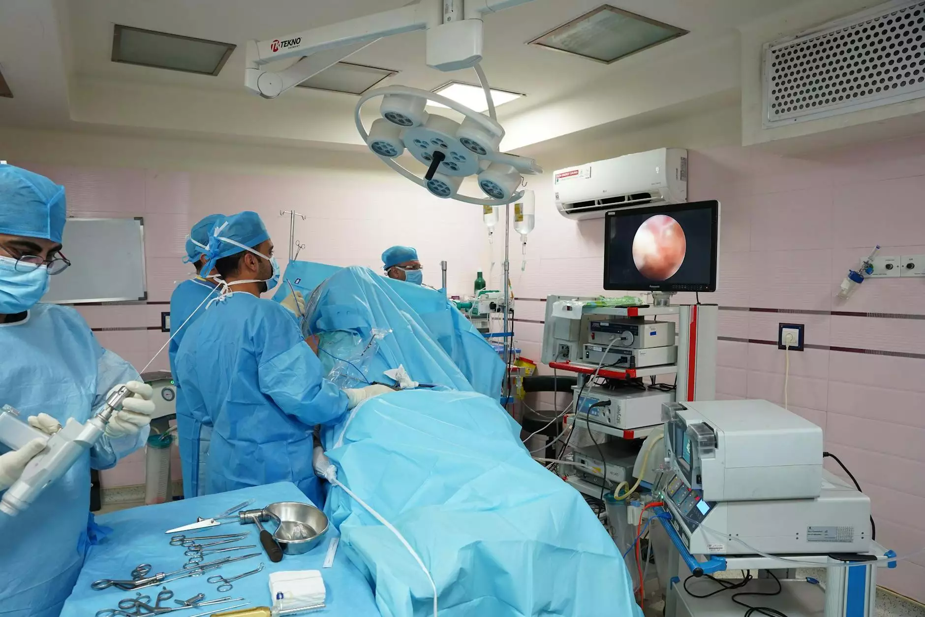
The Pleural Biopsy Procedure: Step-by-Step
The procedure can be performed in several ways, primarily categorized into two types: closed pleural biopsy and open pleural biopsy. Understanding the differences and steps involved in each type ensures patients are adequately prepared.
Closed Pleural Biopsy
The closed pleural biopsy is a minimally invasive procedure often performed in an outpatient setting. Here’s how it typically unfolds:
- Preparation: Before the procedure, patients will usually undergo imaging studies, such as chest X-rays or CT scans, to identify the best site for the biopsy.
- Anesthesia: Local anesthesia is administered to numb the area where the biopsy needle will be inserted.
- Needle Insertion: Using imaging guidance, a long needle is carefully inserted into the pleural space to collect tissue samples.
- Sample Collection: After obtaining the specimen, the needle is withdrawn, and pressure is applied to the site to minimize bleeding.
- Post-Procedure Monitoring: Patients are monitored for a short period to check for complications, such as pneumothorax (collapsed lung).
Open Pleural Biopsy
The open pleural biopsy is more invasive and is conducted in a hospital setting, usually under general anesthesia. Here’s an overview:
- Pre-Operative Assessment: Comprehensive evaluations, including laboratory tests and imaging studies, are performed before the surgery.
- General Anesthesia: The patient is placed under general anesthesia for the procedure.
- Incision: A small cut is made in the chest wall to access the pleura directly.
- Tissue Removal: The surgeon removes a portion of the pleura or surrounding tissue for analysis.
- Closure: The incision is sutured, and the patient is taken to recovery.
Risks and Complications of a Pleural Biopsy
While a pleural biopsy is generally safe, it may present some risks, which patients should discuss with their doctors. Common complications include:
- Pneumothorax: The most significant risk, where air leaks into the pleural space, causing collapse of the lung.
- Bleeding: Minor bleeding occurs but is usually manageable.
- Infection: The procedure can introduce bacteria, leading to infection.
Recovery After a Pleural Biopsy
Recovery from a pleural biopsy varies depending on the type of procedure performed. In most cases:
- Closed Biopsy: Patients can often go home shortly after the procedure, with minimal downtime.
- Open Biopsy: May require a longer hospital stay, and patients will need time to recover from anesthesia and the surgical procedure.
Patients are typically advised to:
- Rest: Avoid strenuous activities for a few days.
- Monitor Symptoms: Watch for signs of complications, such as increased pain, difficulty breathing, or fever.
- Follow-up Care: Schedule follow-up appointments for test results and further evaluation.
Understanding Results from a Pleural Biopsy
The tissue samples obtained during a pleural biopsy are analyzed by a pathologist who examines the cells for abnormalities. Results may take a few days to a week and can provide crucial information regarding:
- Presence of Cancer: Determining if cancerous cells are present.
- Infectious Agents: Identifying microbes responsible for infections.
- Inflammatory Conditions: Determining underlying causes of pleurisy and other inflammatory diseases.
Conclusion: The Importance of the Pleural Biopsy Procedure
In conclusion, the pleural biopsy procedure is a vital tool in the diagnosis and management of numerous pulmonary conditions. For both patients and healthcare providers, understanding the intricacies of this procedure can lead to better health outcomes. By educating oneself about the indications, risks, and recovery expectations, patients can engage actively in their healthcare journey. Always consult with a qualified healthcare provider to determine the best course of action tailored to individual health needs.
For those seeking specialized care, look no further than Neumark Surgery. Our team of experienced doctors is dedicated to providing comprehensive and compassionate care in the field of health and medical services. Visit us online at neumarksurgery.com for more information.